National recognition,
local service.
Board Certified and
Fellowship Trained Radiologists
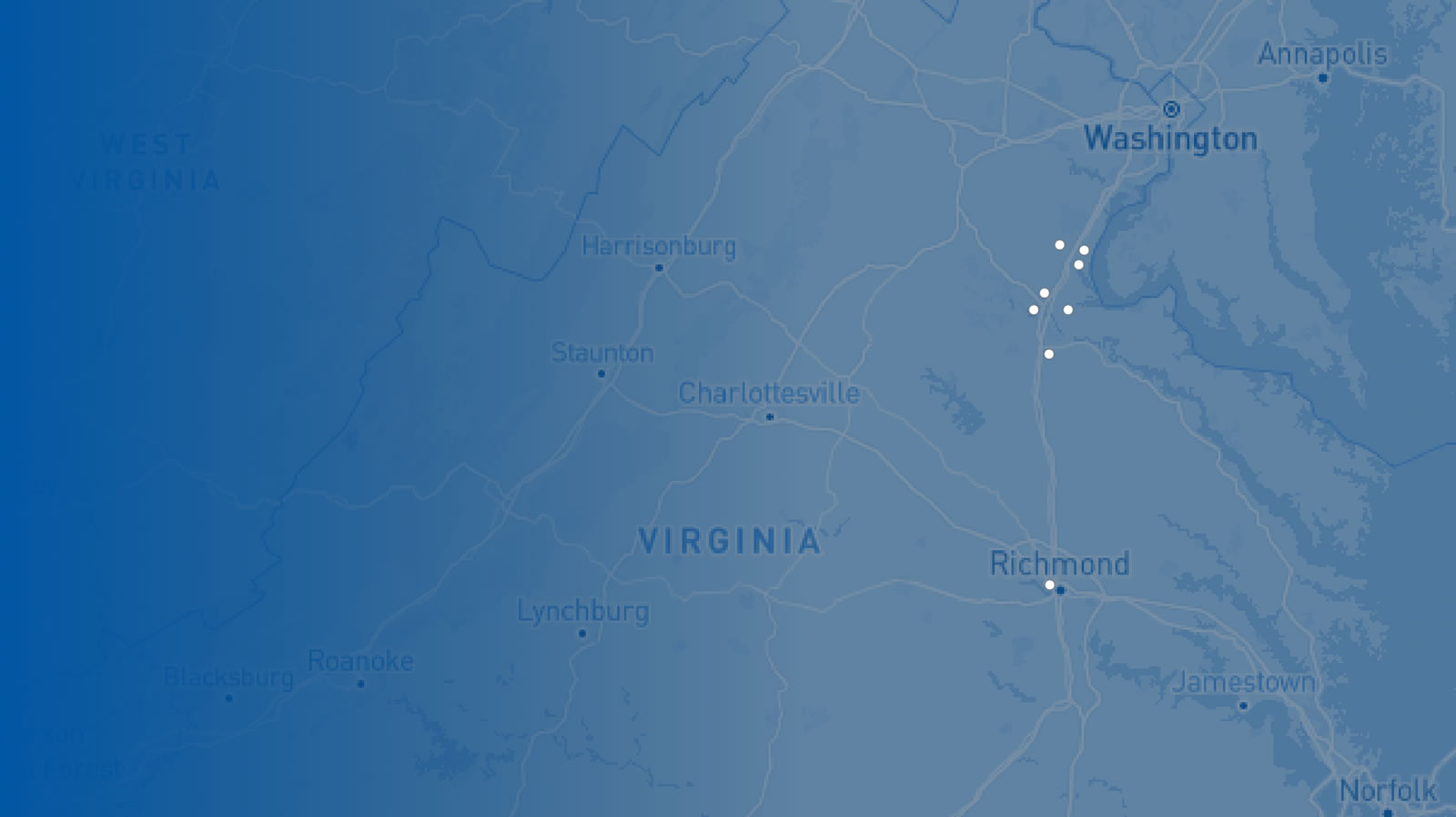
RAF Locations
- Imaging Center for Women
- Imaging Center for Women – North Stafford
- Medical Imaging - Embrey Mill
- Medical Imaging - Fredericksburg
- Medical Imaging - Harrison Crossing
- Medical Imaging - King George
- Medical Imaging - Lee's Hill
- Medical Imaging – North Stafford
- VIVA Fredericksburg
- Administrative Offices
Accreditations
Affiliations


Radiologic Associates of Fredericksburg
10401 Spotsylvania Ave., Suite 200
Fredericksburg, VA 22408
Phone: (540) 361-1000
Fax: (540) 361-7010
Concierge: concierge@rafadmin.com